Marginal mandibular branch of the facial nerve
| Marginal mandibular branch of the facial nerve | |
|---|---|
 Plan of the facial and intermediate nerves and their communication with other nerves. (Labeled at center bottom, second from bottom, as "Mandibular".) | |
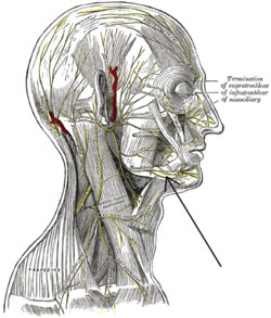 The nerves of the scalp, face, and side of neck. | |
| Details | |
| From | facial nerve |
| Identifiers | |
| Latin | ramus marginalis mandibularis nervi facialis |
| TA | A14.2.01.113 |
| FMA | 53365 |
Anatomical terms of neuroanatomy [edit on Wikidata] | |
The marginal mandibular branch of the facial nerve passes forward beneath the platysma and depressor anguli oris, supplying the muscles of the lower lip and chin, and communicating with the mental branch of the inferior alveolar nerve.
Contents
1 Muscles innervated
2 Clinical significance
3 Additional images
4 References
5 External links
Muscles innervated
The marginal mandibular branch innervates the following muscles:[1]
Depressor labii inferioris - lowers bottom lip down and laterally. Origin: Anterior part of oblique line of mandible. Insertion: Lower lip at midline, blends with muscle from opposite side.
Depressor anguli oris (triangularis) - lowers the corner of the mouth down and laterally. Origin: Oblique line of mandible below canine, premolar, and first molar teeth. Insertion: Skin at the corner of mouth and blending with orbicularis oris.
Mentalis - raises and protrudes lower lip as it wrinkles skin on chin. Origin: Mandible inferior to incisor teeth. Insertion: Skin of chin.
Clinical significance
The marginal mandibular nerve may be injured during surgery in the neck region, especially during excision of the submandibular salivary gland or during neck dissections due to lack of accurate knowledge of variations in the course, branches and relations.
An injury to this nerve during a surgical procedure can distort the expression of the smile as well as other facial expressions.
The marginal mandibular branch of the facial nerve is found superficial to the facial artery and (anterior) facial vein.Thus the facial artery can be used as an important landmark in locating the marginal mandibular nerve during surgical procedures.[2]
Additional images

Lateral head anatomy detail

Lateral head anatomy detail. Neonatal dissection.

References
This article incorporates text in the public domain from page 905 of the 20th edition of Gray's Anatomy (1918)
^ Drake, Richard (2010). Gray's Anatomy of students. Philadelphia: Churchill Livingstone elseveier. pp. 855–866. ISBN 978-0-443-06952-9..mw-parser-output cite.citation{font-style:inherit}.mw-parser-output q{quotes:"""""""'""'"}.mw-parser-output code.cs1-code{color:inherit;background:inherit;border:inherit;padding:inherit}.mw-parser-output .cs1-lock-free a{background:url("//upload.wikimedia.org/wikipedia/commons/thumb/6/65/Lock-green.svg/9px-Lock-green.svg.png")no-repeat;background-position:right .1em center}.mw-parser-output .cs1-lock-limited a,.mw-parser-output .cs1-lock-registration a{background:url("//upload.wikimedia.org/wikipedia/commons/thumb/d/d6/Lock-gray-alt-2.svg/9px-Lock-gray-alt-2.svg.png")no-repeat;background-position:right .1em center}.mw-parser-output .cs1-lock-subscription a{background:url("//upload.wikimedia.org/wikipedia/commons/thumb/a/aa/Lock-red-alt-2.svg/9px-Lock-red-alt-2.svg.png")no-repeat;background-position:right .1em center}.mw-parser-output .cs1-subscription,.mw-parser-output .cs1-registration{color:#555}.mw-parser-output .cs1-subscription span,.mw-parser-output .cs1-registration span{border-bottom:1px dotted;cursor:help}.mw-parser-output .cs1-hidden-error{display:none;font-size:100%}.mw-parser-output .cs1-visible-error{font-size:100%}.mw-parser-output .cs1-subscription,.mw-parser-output .cs1-registration,.mw-parser-output .cs1-format{font-size:95%}.mw-parser-output .cs1-kern-left,.mw-parser-output .cs1-kern-wl-left{padding-left:0.2em}.mw-parser-output .cs1-kern-right,.mw-parser-output .cs1-kern-wl-right{padding-right:0.2em}
^ Batra APS, Mahajan A, Gupta K. Marginal mandibular branch of the facial nerve: An anatomical study. Indian Journal of Plastic Surgery : Official Publication of the Association of Plastic Surgeons of India. 2010;43(1):60-64. doi:10.4103/0970-0358.63968.
External links
Anatomy photo:23:06-0103 at the SUNY Downstate Medical Center - "Branches of Facial Nerve (CN VII)"
lesson4 at The Anatomy Lesson by Wesley Norman (Georgetown University) (parotid3)
cranialnerves at The Anatomy Lesson by Wesley Norman (Georgetown University) (VII)- http://www.dartmouth.edu/~humananatomy/figures/chapter_47/47-5.HTM
This neuroanatomy article is a stub. You can help Wikipedia by expanding it. |


