Condyloid fossa
| Condyloid fossa | |
|---|---|
 Occipital bone. Outer surface. (Condyloid fossa visible but not labeled.) | |
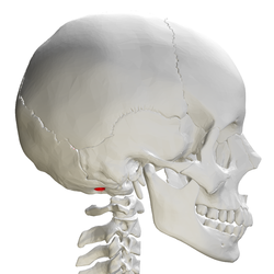 Skull and cervical vertebra. Position of condyloid fossa shown in red. | |
| Details | |
| Identifiers | |
| Latin | Fossa condylaris |
| TA | A02.1.04.017 |
| FMA | 75310 |
Anatomical terms of bone [edit on Wikidata] | |
Behind either condyle of the lateral parts of occipital bone is a depression, the condyloid fossa (or condylar fossa), which receives the posterior margin of the superior facet of the atlas when the head is bent backward; the floor of this fossa is sometimes perforated by the condyloid canal, through which an emissary vein passes from the transverse sinus.
Contents
1 Additional images
2 See also
3 References
4 External links
Additional images
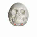
Human skull seen from below. Position of condyloid fossa shown in red.

Skull and cervical vertebra. Position of condyloid fossa shown in red.
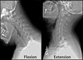
X-ray of cervical spine (neck) in flexion and extension (bending backwards)


See also
- Occipital condyle
- Atlas
References
This article incorporates text in the public domain from page 131 of the 20th edition of Gray's Anatomy (1918)
External links
| Wikimedia Commons has media related to Condyloid fossa. |
- Illustration (#22)
This human musculoskeletal system article is a stub. You can help Wikipedia by expanding it. |


